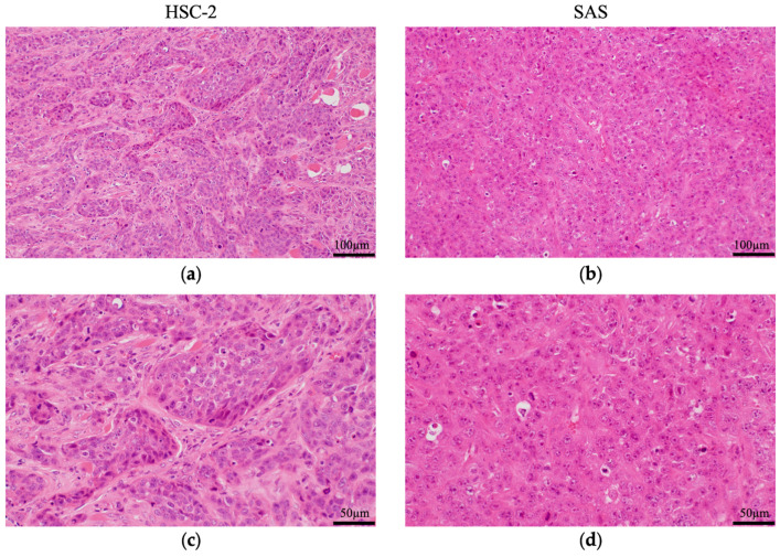
Figure 2.
Pathological analysis of HSC-2 and SAS tumors in mice. Hematoxylin–eosin (HE) staining of resected (a) HSC-2 and (b) SAS tumors. High-magnification images of (c) HSC-2 and (d) SAS tumors. HSC-2 xenografts formed a tumor mass and tumor cells differentiated into basal cell-like and spinous cell-like cells, with fibrous connective tissue and blood vessels in the stroma. SAS xenografts did not form tumor alveoli and proliferated diffusely with necrosis. The tumor cells were poorly differentiated with poor stroma. n = 5.

