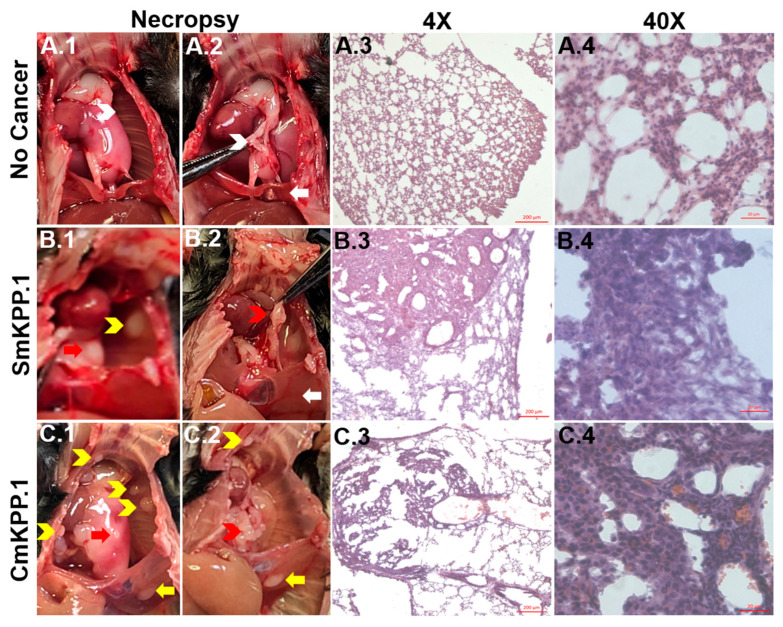
Figure 1.
Visual inspection at necropsy and H&E staining of normal, healthy murine lung tissue and orthotopic tumors generated with monoclonal SmKPP.1 or CmKPP.1 cells. At necropsy, compared to a mouse with no cancer (A.1,A.2)), visual confirmation can be made of primary tumor (left lung, (B.1,C.1), red arrows), metastases to ribs ((B.1,C.1), yellow chevrons), enlarged mediastinal lymph nodes ((B.2,C.2), red chevrons) and metastases to the left hemidiaphragm ((C.1,C.2), yellow arrow). H&E Images at 4× reveal that orthotopic tumors generated by SmKPP.1 (B.3) and CmKPP.1 (C.3) consist of dense sheets of cells surrounded by healthy lung interstitium and alveoli, which normally appears as a delicate, thin, lacy network of cells (A.3). H&E at 40× reveals that both SmKPP.1 (B.4) and CmKPP.1 (C.4) tumors have abundant cytoplasm and have predominantly vesicular nuclei with notable nucleoli compared to normal healthy lung tissue (A.4). All images are 10 μm-thick tissue sections. Scale bars: 200 µm for 4× and 20 µm for 40×.

