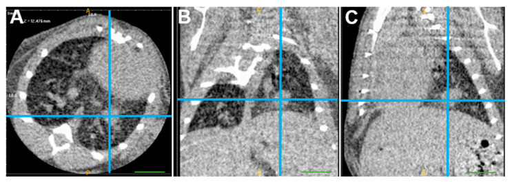
Figure 4.
µCT scan of starting tumor volume of a mouse with an orthotopic tumor generated by 125,000 SmKPP.1 cells. In this representative mouse, one week after injection of SmKPP.1 cells, the left lung tumor is clearly identified (blue crosshairs) in the axial (A), coronal (B), and sagittal (C) planes using ITK-SNAP software, version 3.8.0. Diameters measured in each plane were then applied to a formula for the volume of an oval, the volume of this orthotopic tumor is 6.19 mm3. The scale bar represents 1.0 mm.

