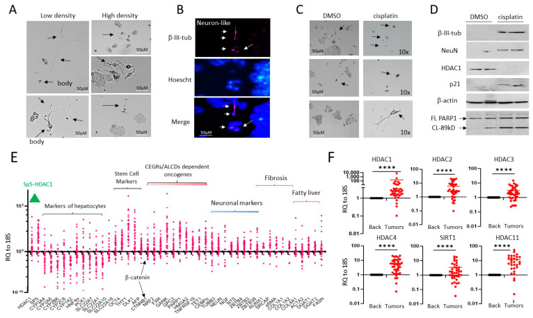
Figure 1.
HepG2 cells contain a small portion of neuron-like cells that are resistant to cisplatin. (A) Images of HepG2 cells plated at high and low density. Arrows show cells that have the shape of neurons. (B) Immunostaining of HepG2 cells with β-III-tubulin. Arrows show positive cells. (C) HepG2 cells with the shape of neurons are resistant to cisplatin treatments. Arrows show cells that have the shape of neurons. (D) Western Blot analysis of proteins isolated from DMSO and cisplatin-treated HepG2 cells. Western blot of PARP1 shows that cisplatin-treated cells express PARP1 band (89 kD), a result of cleavage and an indicator of apoptosis. (E) mRNA expression of several pathways, including HDAC1-Sp5, hepatocyte markers, stem cells, and neuronal markers in a fresh biobank of HBL specimens (n = 42). RQ to18S shows ratios of mRNAs to the 18S RNA. (F) Examination of HDAC family in the biobank of pediatric liver cancers by QRT-PCR. A paired t-test was performed for background and tumor samples—**** denotes a p-value less than 0.0001. The whole gel images of western blots can be found in File S1.

