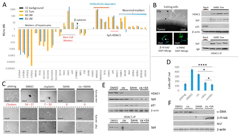
Figure 8.
HDAC inhibition in a lung metastasis-derived cell line increases cisplatin efficacy to eliminate metastatic cells. (A) Characterizations of the cancer pathways in a primary liver tumor and in two lung metastases of a patient who had five total lung metastases. QRT-PCR analysis with a background region (adjacent to tumor), with liver primary tumor (#31), and with two lung metastases (#60 and #81) are shown. (B) Left: An example of exit of LM81 cells from the original Lung Metastasis and staining of the exiting cells with β-III-tubulin and α-SMA. Right, examination of HDAC1-Sp5 p21 pathway in the LM81 and in the LM 81cell line. (C) Treatments of LM81 cells with cisplatin, SAHA, and the combination of cis+SAHA. Red text shows the number of tumor clusters on the plates with low density plating. (D) The number of cells on plates with LM81 cells loaded at high density. * shows p < 0.05, **** shows p < 0.0001. (E) The HDAC1-Sp5 pathway is eliminated by combined treatments with cis+SAHA. The upper part shows levels of HDAC1, Sp5 and p21; the bottom shows HDAC1-IP and Western blot to Sp5. (F) Western blot analysis of proteins isolated from plates treated with DMSO, cisplatin, SAHA and cis+SAHA. The filters were probed for α-SMA, β-III-tubulin and NGF. The whole gel images of western blots can be found in File S1.

