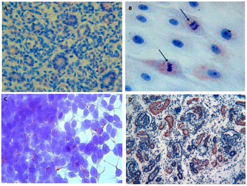
Figure 11.
Tβ4 and Tβ10 in human nephrogenesis. (A) Fetal kidney medullary zone: immunoreactivity for Tβ4 is shown in the cytoplasm of tubular cells. (magnification ×100) (B) PK1 renal cell line. Scattered cultured renal cells immunostained for Tβ4 show a vesicular cytoplasmic staining, which appears stronger in mitotic cells (arrows) (magnification ×630). (C) 293T cell line. Embryonic renal progenitor cells show a peculiar expression pattern for Tβ4. The peptide is mainly expressed in thin cytoplasmic processes, suggesting a role for Tβ4 in intercellular communications. (magnification ×630). (D) Immunostaining for Tβ10 in the fetal kidney is mainly restricted to the proximal and distal tubules. No reactivity for the peptide is observed, in this picture, in the glomerular cells. (magnification ×100).

