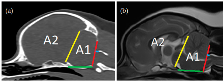
Figure 2.
Cranial fossa measurements: (a) computed tomography, midsagittal plane reconstruction (bone window); (b) magnetic resonance imaging—T2-weighted midsagittal plane. Distance between the os tentorium cerebelli and the dorsum sellae (yellow line); length of the clivus (green line); height of the foramen magnum (‘foramen magnum line’, red line); distance between cranial tip of dorsal arch of the atlas and the foramen magnum line (blue line); area between the yellow line and the red line and osseous structures (caudal cranial fossa area, Area 1); area rostral to the yellow line (rostral and middle cranial fossa area, Area 2).

