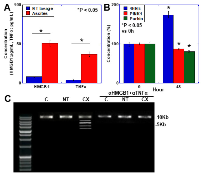
Figure 3.
Mitochondrial changes in the skeletal muscle of a mouse cachexia model. (A) Concentrations of HMGB1 and TNFα in ascites from CX mice used for peritoneal lavage in NT mice. (B) Levels of 4HNE, and protein levels of PINK1 and Parkin in C2C12 cells treated with HMGB1 (50 μg/mL). (C) Mitochondrial DNA alterations in the mouse cachexia model. Mice in the αHMGB1 + αTNFα group were injected with αHMGB1 (0.5 μg/mouse) and αTNFα (0.5 μg/mouse) intraperitoneally three times weekly. Mitochondrial major arc DNA was amplified by PCR. 10 kb, normal-sized band. * Error bars: standard deviation from three independent trials or five mice. Statistical differences were calculated using analysis of variance with the Bonferroni correction. C, control; NT, no tumor; CX, cachexia model; 4HNE, 4-hydroxynonenal; PINK1, PTEN-induced putative kinase 1; αHMGB1, anti-HMGB1 antibody; αTNFα, anti-mouse TNFα antibody; PCR, polymerase chain reaction.

