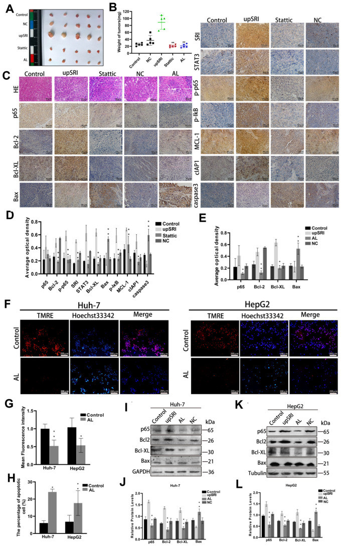
Figure 6.
SRI inhibits cells apoptosis through the NF-κB signaling pathway. (A,B) Tumor volume and weight of orthotopic xenograft models derived from upSRI cells treated with Stattic. Symbols of different colors represented the weight of tumors in different groups. (C,D) Representative IHC images of STAT3, p65, p-p65, p-IκB and apoptosis-related proteins in Stattic treatment. Scale bars = 50 μm. (C,E) Representative IHC images of p65 and apoptosis-related proteins after treatment with AL inhibitor. The expression of proteins were determined by the brown area. (F–H) AL inhibits the fluorescence intensity of TMRE (shown in red) and reduced the sensitivities to SRI-induced apoptosis by Hoechst 33342 (shown in blue) staining assay. Scale bars = 200 μm. (I–L) Apoptosis-related proteins were detected after treated with AL inhibitor by Western blot. * p < 0.05, ** p < 0.01.

