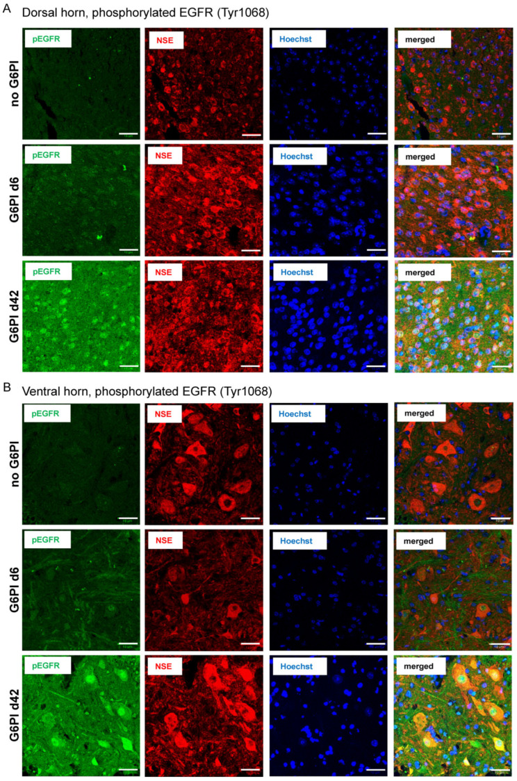
Figure 6.
Localization of pEGFR and colocalization with neuronal markers in the spinal cord of mice in the course of G6PI-induced arthritis. (A) pEGFR (green), NSE (red), nuclear staining (Hoechst 34580, blue), and merged staining of pEGFR and NSE (yellow to orange) in the dorsal horn of mice without inflammation (no G6PI), in mice at day 6 of G6PI-induced arthritis (G6PI d6), and in mice at day 42 of G6PI-induced arthritis (G6PI d42). (B) pEGFR, NSE, Hoechst 34580, and merged staining in the ventral horn of mice without inflammation (no G6PI), in mice at day 6 of G6PI-induced arthritis (G6PI d6), and in mice at day 42 of G6PI-induced arthritis (G6PI d42). Mice were not depleted from Treg cells. Scale bar 10 µm.

