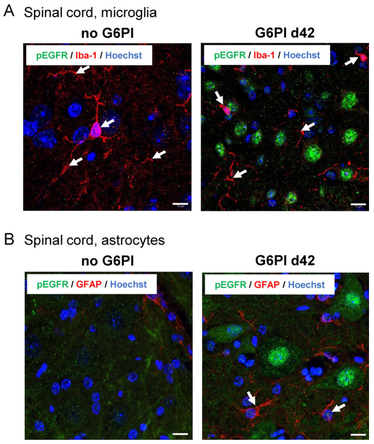
Figure 8.
Representative images of pEGFR labeling in spinal cord microglia and astroglia (n = 3). (A) Spinal cord microglia (Iba-1, red) in the deep dorsal horn of control animals (no G6PI) and at day 42 of G6PI-induced arthritis (G6PI d42). At day 42, pEGFR (green) is not localized in microglia but in neurons. (B) Spinal astroglia in the ventral horn of control animals (no G6PI) and at day 42 of G6PI-induced arthritis (G6PI d42). No GFAP-positive activated astrocytes (red) and no pEGFR (green) in control animals (no GPI), and no pEGFR in GFAP-positive astrocytes at day 42 of G6PI-induced arthritis (G6PI d42). Arrows: examples of positively labeled cells, Scale bar 10 µm.

