Figure 1.
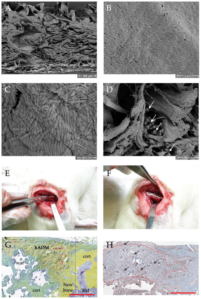
Structure, vascularization and application of hADM in preclinical animal studies [42]. Scanning electron microscopy images of the structural composition at low magnification (A) and evidence of fibrillar collagen at higher magnifications (B,C). Colonization of the hADM with mononuclear cells from the bone marrow (D), arrows mark cells). In (E,F) the application of the hADM in the rat femoral defect model is shown. Movat pentachrome staining of a histological section (G) shows the integration of the membrane (hADM) into newly formed bone tissue (new bone, further abbreviations: cart = cartilage, cort = cortical bone, scaf = scaffold, dashed line shows initial defect border, scale bar = 1 mm). (H) shows the immunohistological evidence of membrane vascularization by specific staining of a-smooth muscle actin. Blood vessels appear brownish and are marked with arrows. The dashed outline indicates the hADM, scale bar = 1 mm.

