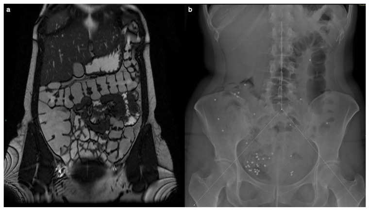
Figure 1.
MR enterography showed no significant alteration two years before surgery, when the patient was already symptomatic. The Coronal T2-weighted image does not show signs of right colon amyloidosis (a). The plain abdominal X-ray is divided into three segments, and radiopaque markers are counted for each segment (b). Measurement of colonic transit time based on radio-opaque markers shows delayed colonic transit time (39/60 markers). The distribution of the markers, with a majority in the right region (31/39), suggests reduced motility at the level of the cecum and ascending colon.

