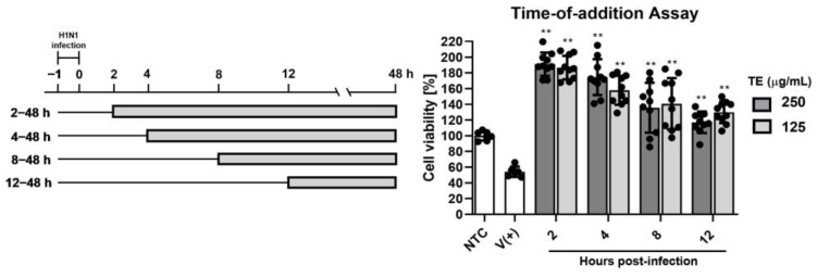
Figure 8.
Time-of-addition assay: MDCK cells were infected with influenza virus (100 TCID50/well) and incubated for 1 h at 37 °C (−1 to 0 h). Virus was removed, and cells were treated with 250 or 125 µg/mL of TE extract starting 2, 4, 8, or 12 h after infection. NTC: non-infected and non-treated control group; V (+): infected and non-treated group; ** p < 0.01 (compared with virus-only control).

