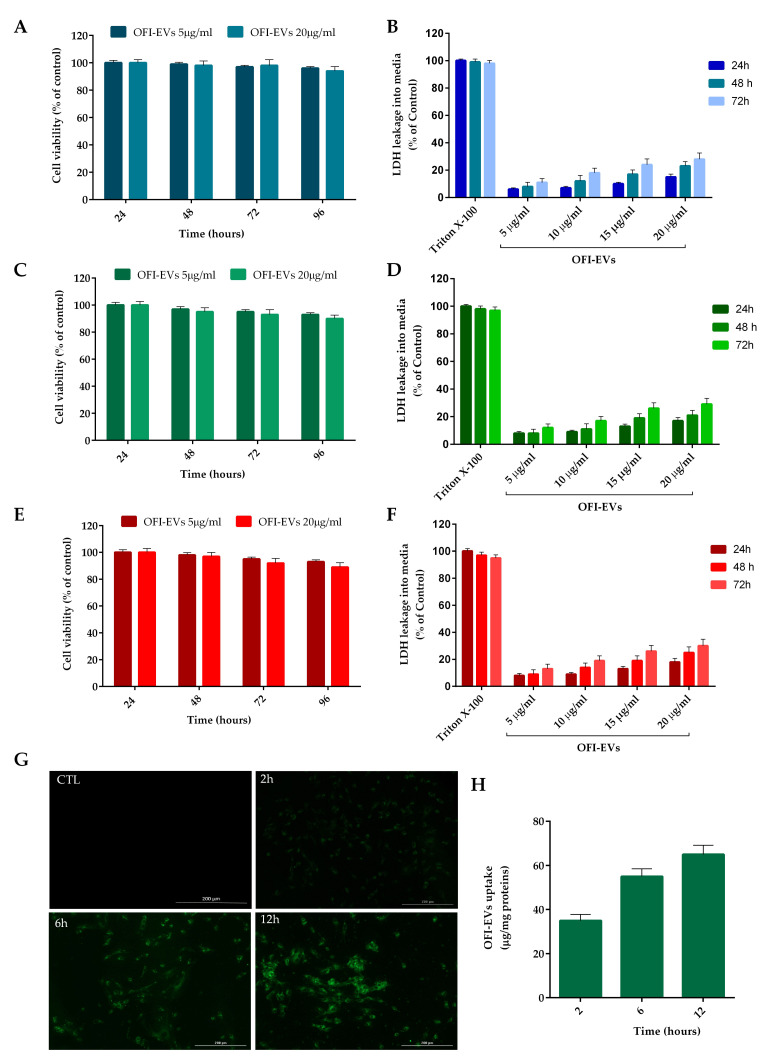
Figure 3.
In vitro biocompatibility and uptake of OFI-EVs. (A) Cytotoxicity was determined in HDFs, (C) HUVECs, and (E) THP-1 cells after 24, 48, and 72 h of incubation with OFI-EVs 5 and 20 µg/mL. A Lactate dehydrogenase (LDH) assay was performed in HDFs (B), HUVECs (D), and THP-1 (F) cells with 5, 10, 15, and 20 µg/mL. Untreated cells were used as controls. (G) EV internalization at different time points. Cells were treated with 20 µg/mL of OFI-EVs dyed with PKH67 (green fluorescent dye). Negative control was without EVs. (H) Semi-quantitative analysis of PKH67-EVs fluorescence intensity in the cytoplasm was evaluated with respect to µg/mg proteins. Data are expressed as the means of three independent experiments ± S.D (n = 3).

