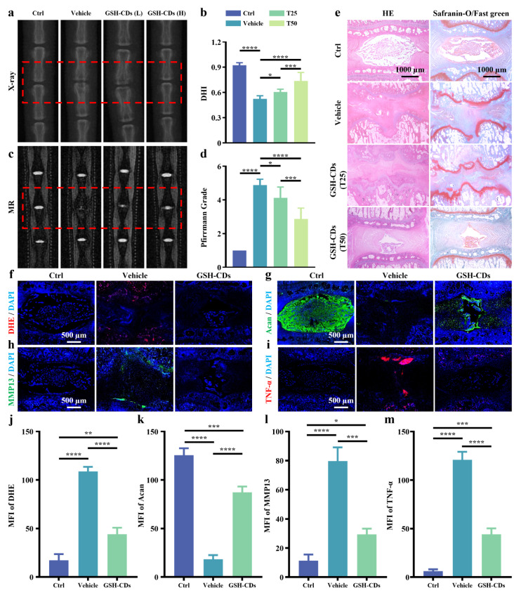
Fig. 6.
Imaging and histological changes in the caudal vertebrae of rats. (a) X-ray images and (b) DHI of the control group, vehicle group, L-GSH-CD group, and H-GSH-CD group. (c) MR images and (d) Pfirrmann grades of the different groups. (e) HE and SOFG staining of different groups (Scale bar = 1000 μm). Images of immunofluorescence fluorescence staining of DHE (f), Acan (g), MMP13 (h), and TNF-α (i) (scale bar = 500 μm) and semiquantitative analysis of the fluorescence intensity of DHE (j), Acan (k), MMP13 (l), and TNF-α (m) in the control group, vehicle group, and GSH-CD group. (* indicates a p value < 0.05, ** indicates a p value < 0.01, *** indicates a p value < 0.001, **** indicates a p value < 0.0001)

