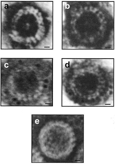
FIG. 3.
Electron micrographs of uranyl acetate-stained RDV particles. Shown are double-shelled VLPs assembled in cells of transgenic rice lines 1 (a) and 2 (b) coexpressing the P3 and P8 structural proteins. Also shown is immunogold probe detection of P8 in VLPs from transgenic rice plant lines 1 (c) and 2 (d). (e) RDV particle purified from RDV-infected rice plants. Bars = 10 nm.

