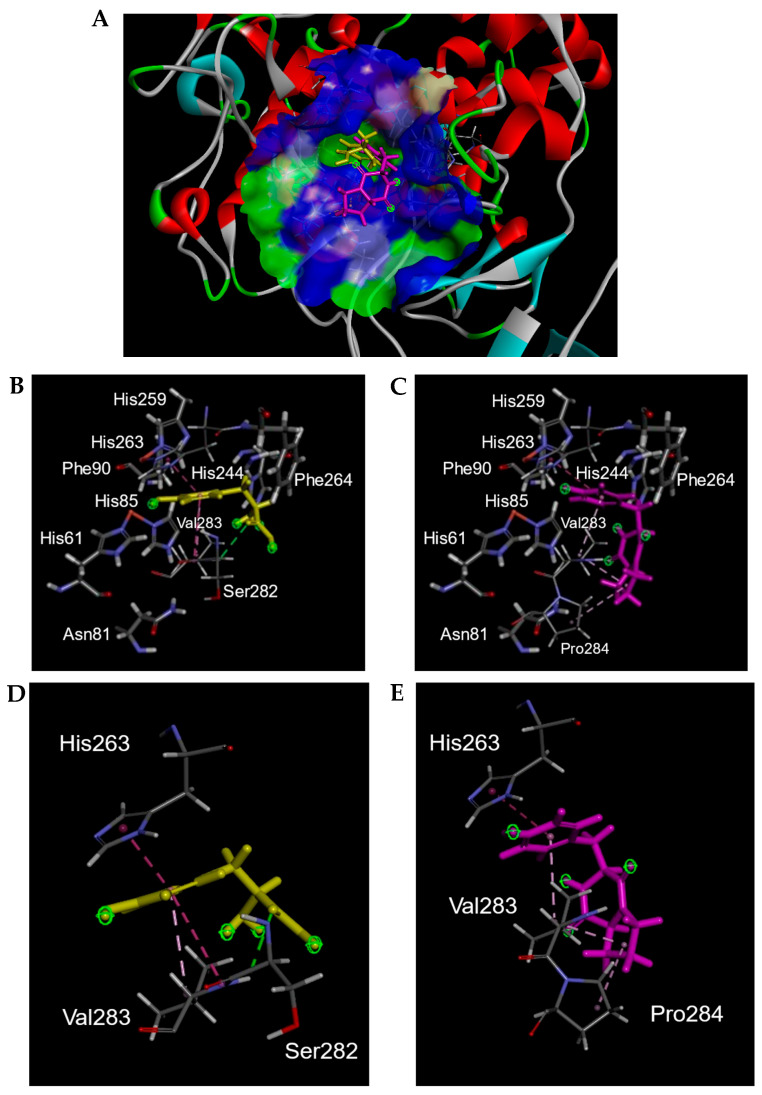
Figure 7.
Three-dimensional (3D) plots of molecular docking diagrams of l-tyrosine and cyclo(l-Pro-l-Tyr) complexes with tyrosinase. (A) These two compounds are indicated with tyrosinase. Yellow: Tyr; light purple: l-Pro-l-Tyr; blue and green areas indicate solvent-accessible surfaces of tyrosinase. Tyrosinase is indicated with red, gray, cyan, and green ribbons. Both l-tyrosine and cyclo(l-Pro-l-Tyr) were positioned in the substrate-binding pocket. Interaction of (B,D) l-tyrosine and (C,E) cyclo(l-Pro-l-Tyr) with tyrosinase in the substrate-binding pocket. (B,C) Amino acids comprising the substrate binding pocket are shown. (D,E) Only amino acids interacting with l-tyrosine or cyclo(l-Pro-l-Tyr) are indicated. Yellow: Tyr; light purple: l-Pro-l-Tyr.

