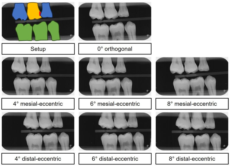
Figure 4.
Digital in vitro bitewing images. Top: color-coded setup—yellow: examination tooth, red: carious lesion, blue: adjacent tooth, green: antagonistic tooth. Below: The mesial-eccentric series shows increased superimposition as the ray path becomes increasingly eccentric in the proximal region of teeth 46 and 47. Conversely, the distal-eccentric series shows increased superimposition as the ray path becomes increasingly eccentric in the interproximal region of teeth 15 and 16.

