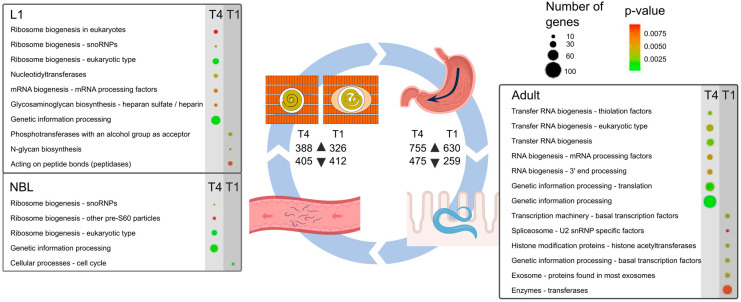
Figure 1.
The diagram represents the Trichinella life cycle, in which first-stage larvae (L1) within striated muscle are ingested and then released in the stomach, progress through to the small intestine, enter the epithelium of the small intestine, develop to fourth-stage larvae and then to the adult stage (within 48 h), copulate, adult males die and adult females lay newborn larvae (NBLs) into lacteals and capillaries, after which individual larvae enter and establish within striated muscle cells. The tables show KEGG BRITE gene family enrichments in NBL, L1, and adult stages between Trichinella pseudospiralis (T4) and T. spiralis (T1). p-values are colour-coded in circles from green to red (low to high); the size of a circle indicates the number of genes linked to an enriched pathway or process. For each species, the numbers of “up-regulated” (▲) and “down-regulated” (▼) transcripts are indicated.

