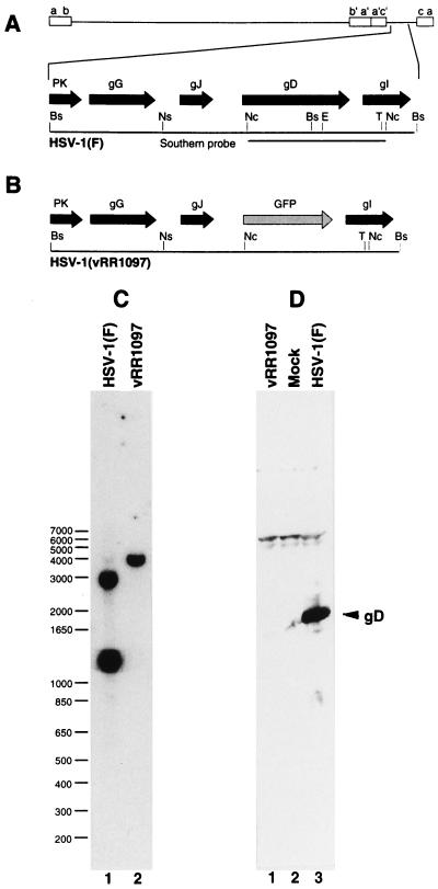
FIG. 3.
Structure of recombinant vRR1097 genome and gD expression in recombinant virus. (A and B) Sequence arrangement of viral genomes of HSV-1(F) (A) and vRR1097 (B) around the gD locus showing the positions of viral open reading frames (solid arrows), GFP insertion (shaded arrow), and probe used for Southern analysis of recombinant virus genome structure. (C) Autoradiographic image of a Southern blot of BstZ17I-digested viral DNAs from cells infected with HSV-1(F) (lane 1) or the gD deletion virus vRR1097 (lane 2). The sizes of migration standards (in kilobase pairs) are indicated to the left of the panel. (D) Photographic image of a Western blot of SDS-polyacrylamide gel electrophoresis-separated proteins from cells either mock infected (lane 2), or infected with HSV-1(F) (lane 3) or vRR1097 (lane 1). The position of the gD signal is indicated by the arrow. Restriction enzyme abbreviations: Bs, BstZ17I; E, EcoNI; Nc, NcoI; Ns, NsiI; T, Tth111I.

