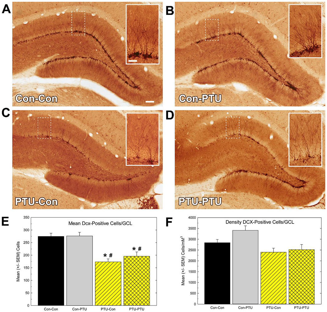
Figure 8.

Immunostaining of neuronal differentiation marker doublecortin (Dcx). (A-D) Low and (B) high magnification (inset) images of Dcx-positive cells in granule cell layer (GCL) of adult hippocampus. (E) Mean (±SEM) Dcx-positive cell number/section. This estimate reflects the average number of cells counted in the GCL of one hemisphere in one section calculated from the mean of all the sections (6-8) assessed for each animal. (F) Mean (± SEM) Dcx-positive cell density. This estimate is the calculated total number of Dcx-positive cells within the hippocampus using stereological methods and normalized to the volume of the GCL (see text). Significant effects in ANOVA were followed by Duncan’s Multiple Range Test for between group comparisons, * different from Con-Con, # different from Con-PTU at p<0.05. Calibration bars 35 and 140 μM.
