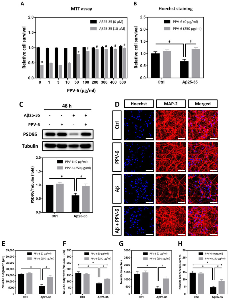
Figure 1.
PPV-6 protects cortical neurons against Aβ25-35 toxicity with restoration of neuronal structure. (A) Primary cortical neurons were exposed to Aβ25-35 (10 μM) with or without PPV-6 at indicated concentrations for 48 h before MTT assay. Mean ± SEM from N = 4. * denotes p < 0.05 compared with control groups without Aβ25-35 treatment; # denotes p < 0.05 compared with Aβ25-35 groups without PPV-6 treatment. (B,C) Cortical neurons were exposed to Aβ25-35 (10 μM) with or without PPV-6 (250 μg/mL) for 48 h before Hoechst staining (B) or western blotting to detect expression of PSD-95 (C). Mean ± SEM from N = 3 in (B) and N = 5 in (C). * and # denote p < 0.05. (D–H) Cortical neurons were treated with or without Aβ25-35 (10 μM) or PPV-6 (250 μg/mL) for 48 h before immunostaining with the MAP-2 antibody (red) to label mature neurons; Hoechst 33258 (blue) served as the counterstaining. Scale bar = 50 μm. Quantitative analyses of total neurite length (in μm), mean neurite length (in μm) per neuron, total numbers of the neurite branch, and mean numbers of the neurite branch per neuron are shown respectively in (E–H). Mean ± SEM from N = 5. All data were analyzed by means of a one-way analysis of variance (ANOVA) followed by a post-hoc Tukey test. *, #, and + all denote p < 0.05.

