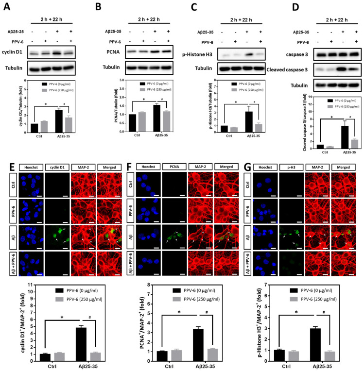
Figure 6.
PPV-6 post-treatment downregulates expression of the cell cycle/apoptosis markers induced by Aβ25-35. (A–D) Primary cortical cultures were exposed to Aβ25-35 (10 μM) for 2 h; this was followed by treatment with PPV-6 (250 μg/mL) for additional 22 h in the absence of Aβ25-35 before detection of cyclin D1 (A), PCNA (B), p-Histone H3 (C), as well as both pro- and cleaved caspase-3 (D) through western blotting. (E–G) Similarly treated cortical cultures were subjected to immunostaining with antibodies against various cell cycle markers (green), including cyclin D1 (E), PCNA (F), and p-Histone H3 (G). The MAP-2 antibody (red) labeled the mature neurons; Hoechst 33258 (blue) served as counterstaining. White arrows denote the mature neurons positively stained with cell cycle markers. Scale bar = 10 μm. Mean ± SEM from N = 4 in (A–D) and N = 6 in (E–G). Data were analyzed by means of a one-way ANOVA followed by a post-hoc Tukey test. * and # denote p < 0.05.

