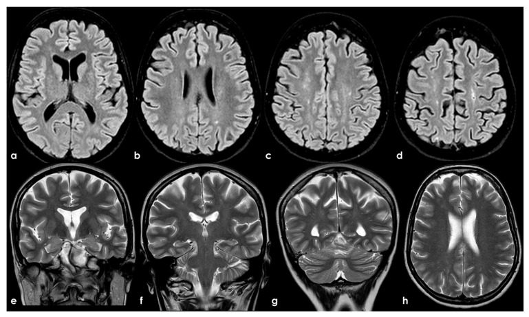
Figure 2.
Brain MRI (axial FLAIR in panels (a–d), coronal T2W in panels (a–g), and axial T2W in panel (h)) showing small punctate white-matter hyperintensities in the centrum semiovale, with a trend to watershed distribution (panel (d)), and a mild increase in enlarged perivascular spaces in the subcortical white matter (panels (e–h)).

