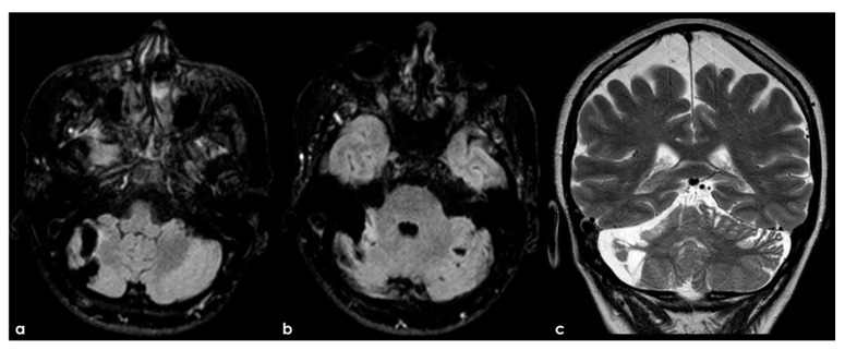
Figure 5.
An example of multifocal cerebellar ischemic lesions in a patient with APS (double positivity). Brain MRI (axial FLAIR in (a,b), coronal T2W sequence in (c) shows the poromalacic evolution of multiple ischemic lesions involving both cerebellar hemispheres (right ≥ left). No causes other than APS were identified in this patients.

