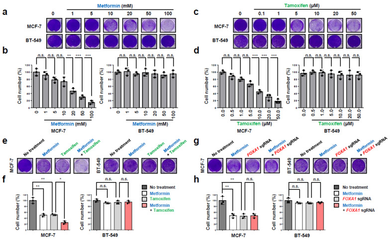
Figure 7.
Metformin and FOXA1 deletion enhance tamoxifen-mediated tumor growth inhibition in hormone-receptor-positive breast cancer (HR+ BC) cells. (a–d) MCF-7 and BT-549 cells were treated with metformin at different concentrations (0, 1, 5, 10, 20, 50, and 100 mM) or tamoxifen at different concentrations (0, 0.1, 1, 5, 10, 20, and 50 μM). After 3 days, the cell viability was measured. Representative image of cells stained with 0.1% crystal violet. The results represent the means (±SE) of three independent experiments performed in triplicate. ** p < 0.01, *** p < 0.001; n.s., non-significant. Statistical comparisons of scatter plots and bar graphs were performed by repeated-measures ANOVA with a multiple-comparisons test. (e–h) MCF-7 and BT-549 cells were treated with metformin (20 mM) or tamoxifen (10 μM) or were infected with FOXA1 sgRNA. After 3 days, the cell viability was measured. Representative image of cells stained with 0.1% crystal violet. The results represent the means (±SE) of three independent experiments performed in triplicate. * p < 0.05, ** p < 0.01; n.s., non-significant. Statistical comparisons of scatter plots and bar graphs were performed by ANOVA with a multiple-comparisons test. The relevant data of (b,d,f,h) can be found in the Supplementary Materials.

