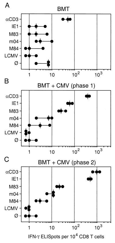
FIG. 3.
Frequencies of IFN-γ-secreting effector T cells in pulmonary infiltrates. Effector cells in the ELISPOT assays were immunomagnetically purified CD8 T cells derived from pulmonary infiltrates. (A) Infiltrates isolated at 4 weeks (phase 1) after BMT with no infection. (B) Infiltrates isolated at 4 weeks (phase 1) during acute infection of the lungs. (C) Infiltrates isolated at 3 months during latent infection of the lungs. The CD3ɛ-redirected ELISPOT assay (Fig. 1) was used to determine the frequency of effector cells irrespective of their antigen specificity. Negative controls included the presentation of an unrelated peptide (LCMV NP aa 118 to 126) by the Ld molecule of P815-B7 cells as well as omission of peptide (∅). Specific peptides of mCMV included IE1 (aa 168 to 176) presented by Ld, M83 presented by Ld, m04 (aa 243 to 251) presented by Dd, and M84 (aa 297 to 305) presented by Kd. Dots represent results from individual assay cultures. Vertical dashes indicate median values.

