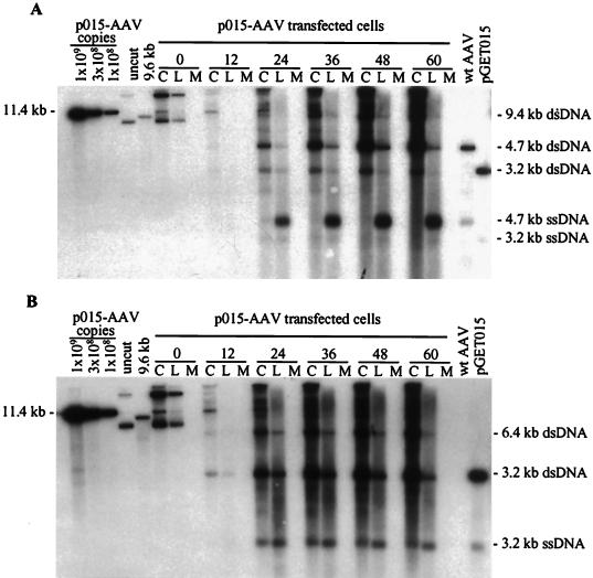
FIG. 2.
Replication of vGET015 is reduced relative to wild type. Ad5dl312-infected 293 cells were processed at the indicated times after transfection with p015-AAV, and 104 cell equivalents of the cell pellet (lanes C), hypotonic lysate (lanes L), and medium (lanes M) were analyzed by Southern blotting using a probe corresponding to the AAV2 terminal repeats (A) or the EGFP gene (B). To estimate the amounts of AAV2 and vGET015 replicative-form DNAs, known amounts of linearized p015-AAV DNA were included in the analysis. p015-AAV (uncut) was used as a marker for input plasmid. A 9.6-kb PpuMI-SwaI fragment of p015-AAV (9.6 kb) was used as a marker for AAV2 dimer-length replicative-form DNA (dsDNA), which is 9.4 kb. A marker for AAV2 monomer-length replicative-form DNA and single-stranded DNA (ssDNA) was prepared by mixing equal amounts of a 4.7-kb PvuII fragment from psub201(−), which was either left untreated or denatured (wt [wild-type] AAV2). Likewise, markers for vGET015 monomer-length replicative-form DNA and single-stranded DNA were prepared using a 3.2-kb AseI-FspI fragment of pGET015 (pGET015). The AseI-FspI fragment (3,170 bp) is slightly larger than predicted size of vGET015 monomer-length replicative-form DNA (3,112 bp).

