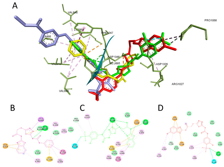
Figure 6.
The most active compounds docked in the binding site of VEGFR-2 PDB:4ASD. (A) Three-dimensional interaction of compound 4a (yellow), 4f (green), 4r (red), and sorafenib(cyan) with the binding site of VEFR-2. (B) Two-dimensional presentation of the interaction of compound 4a. (C) Two-dimensional presentation of the interaction of compound 4f. (D) Two-dimensional presentation of the interaction of compound 4r.

