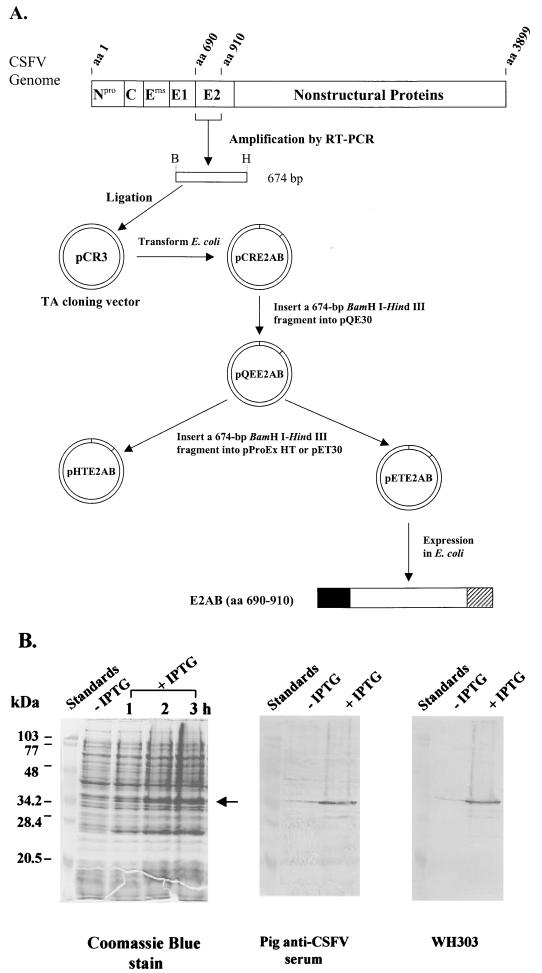
FIG. 1.
(A) Schematic representation of cloning and expression in E. coli of a cDNA fragment coding for an N-terminal portion (aa 690 to 910) of CSFV Alfort/187 E2 protein, designated E2AB. Amino acids are numbered according to the polyprotein derived from the CSFV Alfort/187 genome (27) (GenBank accession number X87939). The restriction endonuclease sites used to clone the cDNA fragment were depicted as follows: B, BamHI; and H, HindIII. Three expression constructs (pQEE2AB, pHTE2AB, and pETE2AB) were created and tested for their ability to express E2AB from T5, trc, and T7 promoters, respectively. E2AB (□) was well expressed only from pETE2AB, yielding the recombinant product with N-terminal (█) and C-terminal ( ) fusions. A six-histidine tag was placed in the N-terminal fusion for affinity purification of the expressed protein. (B) SDS-PAGE and Western blot analysis of the fusion protein expressed from pETE2AB in the absence (−) or presence (+) of IPTG in E. coli. The cells were lysed with the SDS–β-mercaptoethanol sample buffer and heated at 95°C for 5 min before being loaded onto the gel. In all three panels, each lane except the leftmost lane (molecular mass standards) contained total cell proteins from 1 ml of culture with an A590 of 0.2. Left panel, a 3-h time course experiment showing expression of the E2AB fusion protein (indicated by ←) following induction with 1 mM IPTG; middle panel, reactivity of the E2AB fusion protein with polyclonal IgG antibodies of an experimentally CSFV-infected pig serum; right panel, reactivity of the E2AB fusion protein with MAb WH303.

