
Fig. 4.
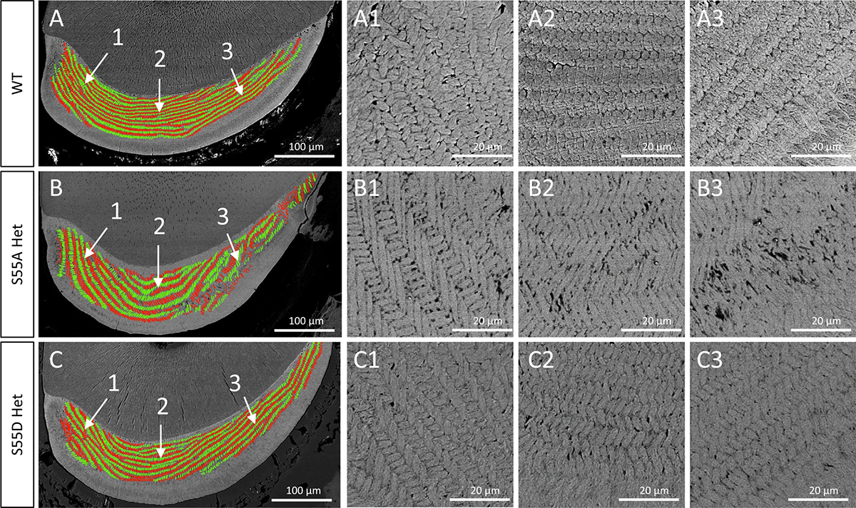
SEM analyses on the transections of lower incisors cut at the crest of alveolar bone near gingival margin of mandibular first molars from 4-week-old mice. In the left panel, enamel rods were color-coded green for rows having a medial tilt and red for rows having a lateral tilt. Sites 1,2,3 represent the medial, middle, and distal regions of incisor section. The panels on right side are higher resolution images (× 800) of the indicated regions in left panels.