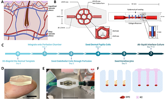
Figure 1.

Description of 3D-SoC biofabrication and model development. (A) Two vascular plexuses that run parallel to the skin’s surface make up the human cutaneous vasculature: the superficial plexus, which is located just under the papillary dermis and surrounds the hair follicles; and the deep plexus, which is located in the lower reticular dermis. (B) Top and side views of CAD drawings of the 3D-SoC model developed in SolidWorks depicting the design and dimensions: a 0.5 mm-diameter horizontally-aligned vascular network (shown in red) surrounding the HF; seven vertically-aligned hair follicle microchannels (shown in blue) with a diameter of 250 µm and a depth of 2 mm, matching the physiological range. (C) Timeline of SoC development showing the significant steps of the protocol. (D) Picture of the 6 mm biopsy sized 3D SoC template biofabricated using the CELLINK Lumen X bioprinter with a photocrosslinkable ink composed of GelMA. Scale bar: 1 cm; (E) picture of the SoC integrated into a 3D-printed perfusion chamber attached on a transparent glass slide; to demonstrate the vascular perfusion, black India ink was perfused through the inlet/outlet ports using a syringe. Scale bar: 2 cm; (F) schematic describing the seeding strategy of DPCs and KCs into the hair follicle microchannels. First, a single cell suspension of DPCs was seeded (shown in red) into the HF microchannels. The cells were given time to settle before the remaining area of the pore was filled with KCs (shown in pink) to achieve a physiologically-relevant conformation found in hair follicle development.
