Figure 3.

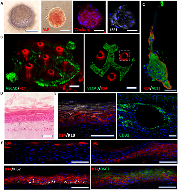
Characterization of the follicular and interfollicular epidermis. (A) Immunofluorescence staining of DPC aggregates at the bottom of the vertical hair follicle microchannels in the SoC model; the expression of alkaline phosphatase (ALP), versican, and LEF1 is associated with functional hair-inductive DPCs. (B) The anticipated spatial distribution of cell types in the printed vascular plexus and hair follicle channels is demonstrated by staining for endothelial cell marker, VECAD (green), and basal epidermal/hair follicle marker, Keratin 14 (K14; red). (C) The proper conformation of the follicular component is further demonstrated by the expression of the outer and inner root sheath markers, K14 and AE13, respectively. (D) H&E of the 3D-SoC showing the interfollicular epidermal compartment and the full development of all epidermal layers. (E) Representative immunofluorescence staining of whole-mount tissues (orthogonal views) showing K10 and K14 expression patterns (left), and CD31 staining to display endothelial cell coverage of the perfusable vasculature (right). (F) Tissue sections showing proper epidermal maturation with basal expression of K14 and Ki67, suprabasal involucrin (IVL) and desmoglein 1 (DSG1), and loricrin enhancing the terminally differentiated layer. The presence of Ki67+ proliferative keratinocytes indicate the homeostatic state of the epidermis and its self-renewal capability. Scale bars: 100 µm for (A), (C), (D)–(F); 250 µm for (B).
