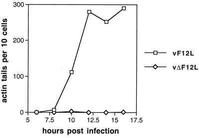
FIG. 5.
Graph showing the number of virus-tipped actin tails in infected cells at different times p.i. BS-C-1 cells were infected with vF12L, vΔF12L, or vF12L-rev at 0.1 PFU/cell and at the indicated times p.i. were stained with MAb AB1.1 to reveal all VV particles and with TRITC-phalloidin to detect F-actin. Samples were analyzed by confocal microscopy and reconstructed z-series of images of different sections through the cell were examined. The number of virus-tipped actin tails was counted for each of 10 infected cells (as shown by reactivity with AB1.1), and the average number is shown. The number of virus-tipped actin tails in cells infected with vF12L and vF12L-rev were indistinguishable, and data are shown for only vF12L compared with vΔF12L.

