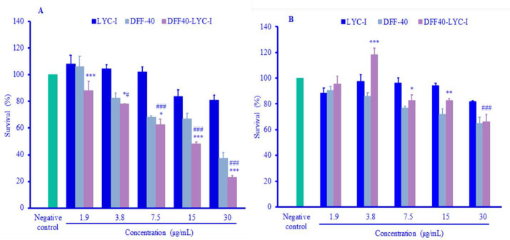
Fig. 5.
Cytotoxicity assays of DFF40, LYC-I and DFF40-LYC-I against different cell lines after 48 h of incubation. (A) HeLa cell line; (B) HUVEC cell line. Data were presented as mean ± SD (n=3). *P . 0.05, **P . 0.01, and ***P . 0.001 represent significant differences than DFF40 in the same concentrations. #P . 0.05 and ###P . 0.001 represent significant differences versus negative control. LYC-I, lycosin-I; DFF, DNA fragmentation factor.

