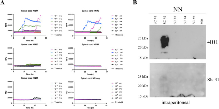
Fig 5. RT-QuIC and western blot analysis of spinal cord homogenates of Prnp.Cer.138NN mice inoculated i.p. with M-NO3.
(A) Ten percent spinal cord homogenates were subjected to serial dilutions from 2 x 10⁻¹ to 2 x 10⁻⁶ in RT-QuIC seed dilution buffer. Samples were considered positive when a minimum of two out of four wells crossed the threshold relative fluorescence unit (RFU). Threshold is the average RFU of all negative control reactions plus five times their standard deviation. Negative control was a naïve spinal cord homogenate of a Prnp.Cer.138NN mouse. The y-axis represents the RFU, and the x-axis represents time in hours (h). Mouse recombinant PrP was used as substrate for the RT-QuIC reactions. (B) Spinal cord homogenates were digested with 50 μg/ml of PK and subjected to western blot analysis using anti-PrP antibodies 4H11 (1:500) and Sha31 (1:10,000); see also overexposed western blot in Fig G in S1 Text).

