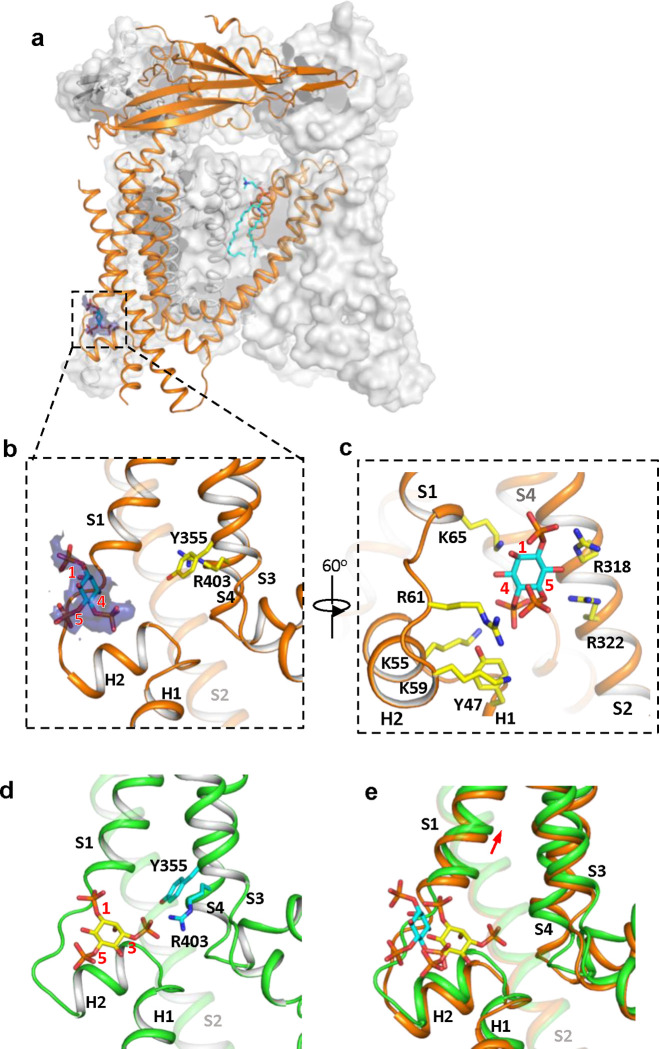
Figure 3. Structure of TRPML1 in complex with PI(4,5)P2.
(a) Overall structure of PI(4,5)P2-bound TRPML1 with the front subunit shown in orange cartoon and the rest shown as grey surface representation. Density for PI(4,5)P2 head group is shown in blue surface. (b) Zoomed-in view of the PI(4,5)P2-binding pocket with the density of its IP3 head group shown in blue surface. (c) Zoomed-in view of the PI(4,5)P2-binding pocket with side chains of IP3-interacting residues shown as yellow sticks. (d) Zoomed-in view of the IP3 position in the PI(3,5)P2-bound open TRPML1 structure. The C3 phosphate group directly interacts with Y355 and R403. (e) Comparison of the head group positions in PI(3,5)P2-bound open (green) and PI(4,5)P2-bound closed (orange) structures. The inositol rings PI(3,5)P2 and PI(4,5)P2 are colored yellow and cyan, respectively. The red arrow marks the upward movement of S1 from closed to open conformation.

