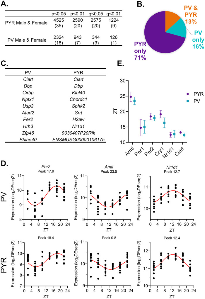
Figure 2. While pyramidal cells have greater transcript rhythmicity, the molecular clock is in-phase across cell types.
(A) The number of rhythmic transcripts at different significance cutoffs in pyramidal and PV cells. The percentage of transcripts that are rhythmic are displayed in parentheses. Pyramidal cells show the most rhythmic transcripts at all cutoffs. (B) The percentage of rhythmic transcripts (p<0.01) present in each cell type as a proportion of all rhythmic transcripts detected. Rhythmic transcripts are largely different between PV and pyramidal cells. (C) The top 10 rhythmic transcripts (by p-value) in PV and pyramidal cells include multiple known circadian genes. (D) Rhythmicity patterns of core clock genes in PV cells (top) and pyramidal cells (bottom). Each point represents a subject, with time of death (ZT) on the X axis and log2DESeq2 normalized expression on the Y axis. The fitted sinusoidal curve is shown in red. (E) The calculated peak time (ZT) and 95% confidence intervals of conserved canonical circadian transcripts, indicating that the core clock is in-phase across cell types. ZT=Zeitgeber time, PYR=pyramidal cells, PV=parvalbumin cells

