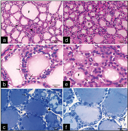
Figure 2.

Photomicrographs of sections of Groups III A1 (a-c) and A2 (d-f) showing: (a) Areas of cellular infiltration (arrowhead). Some follicles (F) appear distended with colloid and lined by flat follicular cells, microcystic follicles (arrows) appear with absent or scanty amount of colloid (H and E ×400) (b and c) some follicular cells with vacuolated cytoplasm (arrows) and irregular and discontinuous basement membrane (arrowhead) (H and E ×1000, toluidine blue, ×1000). (d) Congested blood vessels (arrow) (H&E, x400) (e) few follicular cells with cytoplasmic vacuolations (arrow) (H&E x1000). (f) follicular cells with rounded basophilic nuclei with irregular basement membrane (arrows) (Toluidine blue X1000)
