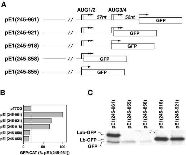
FIG. 7.
Sequence between the two pairs of AUG codons is important for ERAV IRES activity. (A) Diagram of plasmids containing 3′ truncations of the ERAV IRES, highlighting the positions of the AUG codons (arrows). Note that in these plasmids there is no additional sequence between the GFP AUG and the ERAV sequence. (B) GFP-to-CAT ratios in VTF7-3 infected BHK-21 cells (T7 expressing) following transfection with the plasmids named on the left. GFP was measured by FACS analysis of whole cells, while CAT enzyme activity was determined from cell extracts as detailed in Materials and Methods. Values are expressed as percentages of the  ratio determined for pE1(245–961). The vertical line indicates the value obtained for the GFP-negative control plasmid pT7CG. (C) RIP analysis of vTF7-3-infected BHK-21 cell extracts following transfection with the plasmids diagramed in panel A and metabolic labeling with [35S] methionine. Extracts were immune precipitated with GFP antibody-coated protein A beads. Labeling for the pE1(245–961) lane is on the left.

