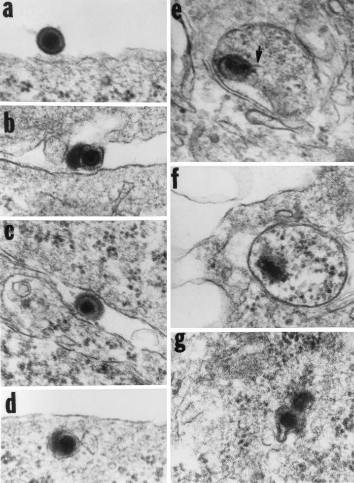
FIG. 4.
Electron micrographs of thin sections of cells exposed to 100 PFU equivalents of gD−/−. SK-N-SH cells were exposed to 100 PFU equivalents per cell and incubated for 30 min.. They were then harvested, fixed, sectioned, and stained for electron microscopy as described elsewhere (4). The arrow in panel e points to a structure typical of DNA streaming out of damaged capsids.

