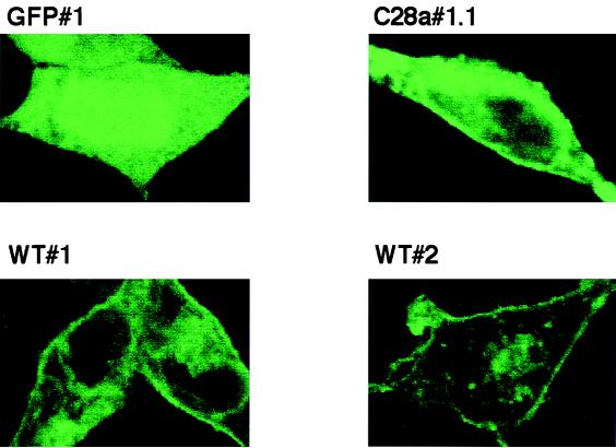
FIG. 3.
Confocal microscopy of selected cell lines GFP#1, WT#1, WT#2, and C28a#1.1. Clonal cell lines stably expressing HN-GFP were plated on collagen coated slides and allowed to attach. Monolayers were fixed using formaldehyde. Cells were visualized under confocal microscopy. All pictures were taken using the same aperture, time of exposure, laser intensity, and magnification. The HN-GFP fusion proteins localize to the plasma membrane. As a control, cells expressing the GFP protein alone show only cytoplasmic fluorescence.

