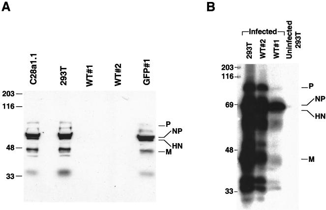
FIG. 6.
Western blot analysis with anti-HPF3 serum of cell lines infected with HPF3. (A) Monolayers of C28a#1.1, 293T, WT#1, WT#2, and GFP#1 cells were infected with HPF3 at an MOI of 0.1 PFU/cell. Cell extracts were collected 24 h after infection. Equal amounts of protein (20 μg) were loaded per lane and analyzed by SDS-PAGE. Immunoblotting was performed with anti-HPF3 serum. The lysates are from the infected cells indicated above the lanes. (B) Monolayers of 293T, WT#2, and WT#1 cells were infected with HPF3 at an MOI of 0.1. Cell extracts were collected 72 h after infection. Equal amounts of protein (20 μg) were loaded per lane and analyzed by SDS-PAGE. Immunoblotting was performed with anti-HPF3 serum. The lysates are from the infected cells indicated above the lanes.

