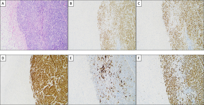
Fig. 2.
Histological and immunohistochemical features of a neuroendocrine carcinoma metastasis to the adrenal gland. A Routine hematoxylin and eosin staining depicting the normal adrenal cortex (left) and a metastatic neuroendocrine carcinoma (NEC) (right). The tumor is arranged in ribbons and solid sheets, with numerous mitotic figures and focal tumor necrosis. B The second-generation neuroendocrine marker ISLET1 is diffusely positive in tumor cells, as was first-generation marker synaptophysin (not shown). C INSM1 is also clearly positive, thus verifying the neuroendocrine nature of this lesion. D Diffuse TTF1 expression in the tumor cells. E CK7 is focally expressed within the tumor. The TTF1 and CK7 stains favored a pulmonary origin. F The Ki-67 labeling index is 80%

