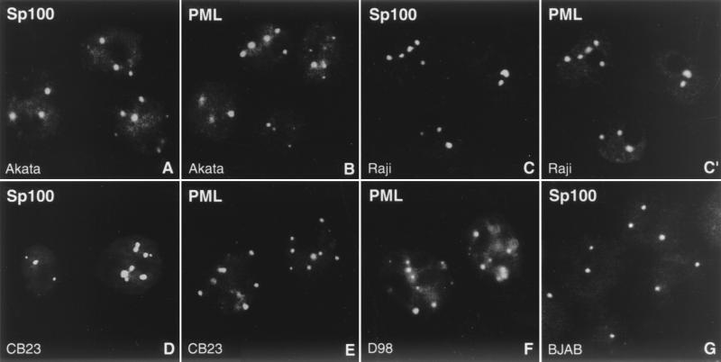
FIG. 1.
ND10 are not disrupted during latency. The EBV-positive Burkitt's lymphoma cell lines Akata (A and B) and Raji (C and C′), the latently EBV-infected lymphoblastoid cell line CB23 (D and E), and the EBV-positive fusion cell line D98/HR1 (D98; F) possess intact ND10, as revealed by immunohistochemistry with antibodies against Sp100 (A, C, D, and G) and PML (B, C′, E, and F). C and C′ shows double labeling to demonstrate colocalization of both proteins. As a control, ND10 are visualized in the EBV-negative Burkitt's lymphoma cell line BJAB by staining for Sp100 (G).

