FIG. 3.
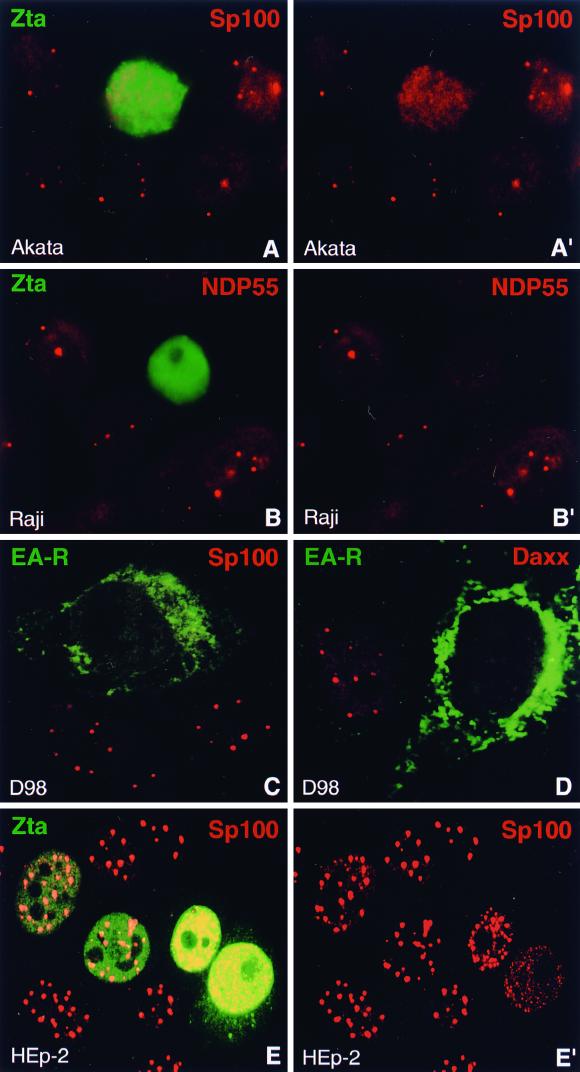
Lytic activation of EBV disperses the ND10 proteins Sp100, NDP55, and Daxx. (A and A′) Lytic activation of EBV was induced in Akata cells and monitored by staining for the immediate-early protein Zta. Sp100 is no longer concentrated in ND10 in Zta-expressing cells. A′, Sp100 staining only. (B and B′) Lytic activation of EBV in Raji cells, detected by staining of Zta, induces the dispersion of the ND10 protein NDP55. B′, NDP55 staining only. (C) D98/HR1 cells were Zta transfected to induce lytic activation of EBV and stained for the viral protein EA-R and Sp100. EA-R, which is localized in the cytoplasm, allows the identification of cells in which EBV has entered the lytic cycle. Sp100 is no longer concentrated in ND10 after lytic activation. (D) Like Sp100 and NDP55, the ND10 protein Daxx disperses in EA-R-positive D98/HR1 cells. (E and E′) EBV-free HEp-2 cells were transfected with a Zta-expressing plasmid and stained with antibodies against Zta and Sp100. ND10 remain intact at Zta concentrations comparable to those observed after lytic activation, but Sp100 starts to disperse when Zta is expressed at very high levels (cells on right). Note the deposition of Zta in the cytoplasm in the lower right cell. E′, Sp100 staining only.

