
Figure 2. Injection of DA increases myeloid cell numbers during the inflammation phase of acute skin wound healing.
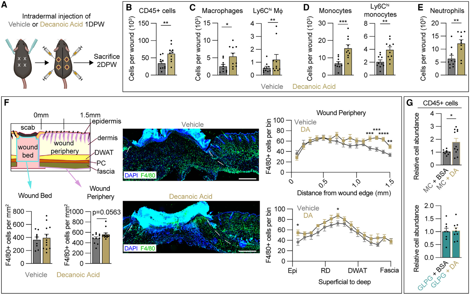
(A) Schematic of decanoic acid (DA) treatment paradigm.
(B–E) Quantification from flow cytometry analysis of Zombie− (B) CD45+ cells, (C) total wound macrophages and Ly6Chi macrophages, (D) total monocytes and Ly6Chi monocytes, and (E) neutrophils 2 DPW (n = 10 wounds from 5 male mice per condition).
(F) Spatial analysis of tissues immunostained for F4/80 to determine macrophage numbers in the wound bed (WB) and wound periphery (WP) of vehicle- and DA-treated animals. Dashed white lines delineate wound edges. Scale bars, 500 μm.
(G) Quantification from flow cytometry analysis of SYTOX− CD45+ immune cells in methyl cellulose- (MC; top) or GLPG1205 (bottom)-treated mice injected intradermally at the WP with vehicle (BSA) or DA (n ≥ 11 wounds for each condition in males).
Error bars indicate mean ± SEM. Significance was determined using a two-tailed Student’s t test. *p < 0.05, **p < 0.01, ***p < 0.001, and ****p < 0.0001. M4, macrophage; DPW, days post-wounding; Epi, epidermis; RD, reticular dermis; DWAT, dermal white adipose tissue.