
Figure 3. Inhibition of GPR84 signaling delays epidermal repair during skin wound healing.
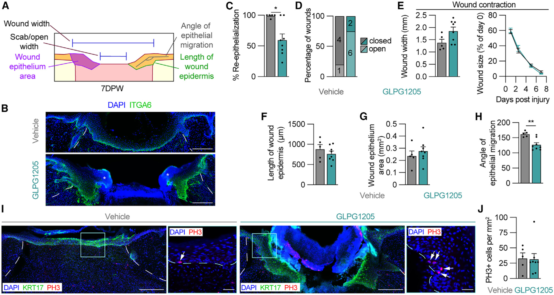
(A) Schematic of measurements used to assess the epithelium of wounds 7 DPW.
(B) ITGA6 immunostained tissue sections from the center of wounds obtained from vehicle- and GLPG1205-treated mice. Asterisks indicate scab.
(C–H) Quantification of parameters that assess epithelial repair including (C) WB re-epithelialization, (D) percentage of open versus closed wounds, (E) wound width and wound size, (F) length of wound epidermis, (G) wound epithelium area, and (H) the angle of epithelial migration. The angle of epithelial migration was measured on both sides of each section analyzed.
(I) Tissue immunostained for KRT17 and PH3. High-magnification images show PH3 in the epithelium. Arrows point to PH3+ nuclei.
(J) Quantification of PH3+ cells per area of wound epithelium (n ≥ 5 wounds from ≥3 male mice per condition). Dashed white lines delineate wound edges, and dashed blue lines delineate epithelium. Scale bars, 250 μm in composite images and 25 μm in high-magnification images. Error bars indicate mean ± SEM. Significance was determined using a two-tailed Student’s t test for all analyses except (D), where a Fisher’s exact test was used. *p < 0.05 and **p < 0.01. KRT17, keratin 17; PH3, phospho-histone H3.