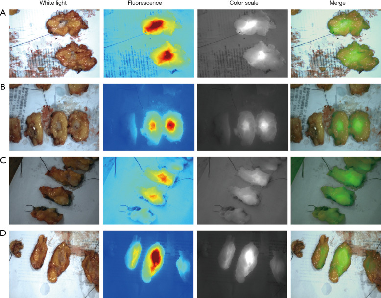
Figure 1.
Fluorescence of dissected tumor. NIF detection of isolated tumors in white light, fluorescence, color scale and merger modes. ICG can be found accumulating in the tumor tissue after sectioning the specimen, which is different from the paracancerous tissue. (A) Patient was injected 4 hours prior to surgery at a dose of 0.5 mg/kg. The tumor size was 1.5 cm. (B) Patient was injected 3 hours prior to surgery at a dose of 0.5 mg/kg with a tumor size of 1.8 cm. (C) Patient was injected 8 hours prior to surgery at a dose of 1 mg/kg with a tumor size of 2.3 cm. (D) Patient was injected 2 hours prior to surgery at a dose of 0.5 mg/kg with a tumor size of 1.9 cm. NIF, near-infrared fluorescence; ICG, indocyanine green.

